Tissue Diagnostic Is Beneficial For Cancer Diagnosis and Developing Treatments for Targeted Therapies
 |
| Tissue diagnostic |
Tissue diagnostic
is a type of histology that uses tissue from organs and tumors to diagnose
diseases. This field of medicine is also known as histology, anatomic
pathology, or cell and tissue diagnostics.
A tissue
diagnostic is an examination of a suspicious tumor. It is an important part
of diagnosing cancer. Tissue samples can be obtained by cutting through the
skin or a soft tissue such as the lining of the intestines (endoscopy). Once
removed, the tissue is sent to a laboratory or department of pathology to be
examined by a histopathologist.
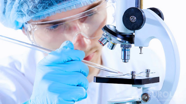
For examining the
sample, a pathologist must first cut it into very small pieces. This process
makes it possible to see the tissue under a microscope. To make the sample more
solid, a substance called formalin is used. Once the tissue has been
stabilized, it is then cut into sections that can be seen under the microscope.
Histopathologists
then analyze the tissue samples to find out if they are malignant. This
information is crucial for treating the patient, as well as for researchers to
understand how different kinds of cancers respond to treatment.
Several methods
are used to detect cancers in tissue samples, including immunohistochemistry
and in-situ hybridisation stains. These stains allow the pathologist to find
specific cells in the tissue, so they can identify the type of cancer and
whether it has spread.
Another method is
to take a small sample of tissue from the tumor, and then place it in a special
dye. The dye then absorbs light and scatters it into a pattern that is visible
under a microscope. The image is then shown to a doctor who can tell what kind
of cancer the tissue sample represents and, if it is malignant.
A second way to
detect cancer is by taking x-rays. This test creates a picture of the inside of
the body, and it can be used to detect cancer in bone sarcomas. However, it is
not as helpful in diagnosing soft-tissue sarcomas.
The most common
method for diagnosing a cancer is by examining the tissue itself. Tissue is
then removed and sent to a lab to be looked at under a microscope by a doctor
who is trained in pathology.
Hoffmann La Roche declared the launch
of next-generation VENTANA DP 600 slide scanner in June 2022. This scanner has
high quality image of stained histology slides and is easy to use.


Comments
Post a Comment